The Rise of 3D Imaging in Modern Dentistry
Introduction to 3D imaging technology in dentistry
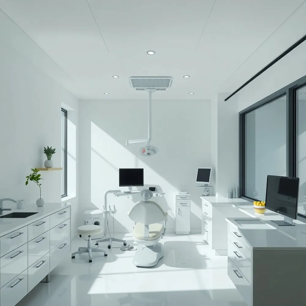
Modern dentistry has been transformed by 3D imaging technologies, offering detailed, three-dimensional views of teeth, gums, bones, and surrounding structures. These advanced imaging tools replace traditional two-dimensional X-rays with comprehensive 3D models that improve the visualization of oral anatomy.
Importance of 3D imaging for diagnostic accuracy and patient care
3D imaging enhances diagnostic precision by revealing hidden fractures, root configurations, bone density, and nerve pathways that are not visible with 2D images. This accuracy enables dentists to tailor treatment plans more effectively, reducing risks during procedures such as implants, orthodontics, and surgery. Patients benefit from faster, less invasive exams combined with improved communication and understanding of their dental health.
Overview of Cone Beam Computed Tomography (CBCT) and digital scans
Cone Beam Computed Tomography (CBCT) stands out in 3D dental imaging for its ability to quickly produce high-resolution scans with minimal radiation exposure. CBCT provides a full 360-degree view in a single painless scan, ideal for implant planning, endodontic treatment, and surgical guidance. Complementary digital scans, like intraoral scanners, generate precise digital impressions, streamlining restorative and orthodontic workflows.
Together, these technologies offer dentists enhanced tools to deliver personalized, efficient, and safe dental care for patients of all ages.
Key Facts on 3D Imaging in Dental Care
- 3D imaging, especially CBCT, provides highly detailed, three-dimensional images that enhance dental implant planning.
- CBCT scans identify critical anatomical structures like the inferior alveolar nerve and the maxillary sinus to reduce surgical risks.
- Digital planning software enables virtual implant placement, allowing customization of implant angles, depths, and sizes.
- 3D-printed surgical guides are created from virtual plans to ensure precise implant placement during surgery.
- Utilizing 3D imaging increases surgical accuracy, safety, and predictability, leading to improved long-term success rates.
- In orthodontics, CBCT combined with intraoral and facial scans creates a comprehensive 3D model for personalized treatment planning.
- 3D visualization aids in designing custom braces, aligners, and retainers, improving fit and comfort.
- Superimposition and deviation software enable monitoring of morphological changes and progress tracking during orthodontic treatment.
- In oral surgery and endodontics, CBCT improves diagnosis precision by revealing complex root anatomies and hidden lesions.
- CBCT-based virtual surgical planning reduces invasive procedures, shortens recovery times, and enhances treatment safety.
1. Enhancing Dental Implant Planning with 3D Imaging
![]()
How does 3D imaging improve dental implant planning?
Cone Beam Computed Tomography (CBCT) has transformed dental implant planning by providing highly detailed, three-dimensional images of a patient’s jawbone and surrounding anatomy. This advanced imaging allows dentists to precisely assess bone height, width, and contour, which are crucial factors for the stability and success of dental implants.
What anatomical details can CBCT scans reveal?
CBCT scans enable clinicians to identify critical anatomical structures such as the inferior alveolar nerve in the lower jaw and the maxillary sinus in the upper jaw. Recognizing these vital features helps avoid nerve damage and sinus perforation, substantially reducing surgical risks and complications during implant placement.
How is digital technology integrated into the implant planning process?
Using CBCT data, dentists employ specialized digital planning software to perform virtual implant placement. This software allows for testing various implant angles, depths, and sizes in a virtual environment, helping tailor the procedure to the patient’s unique anatomy and restorative goals.
What role do 3D-printed surgical guides play?
From the virtual plan, surgical guides can be 3D printed with remarkable precision. These guides fit snugly onto the patient’s teeth or gums during surgery, ensuring that implants are placed exactly as planned. This increases procedural accuracy and safety, especially in complex cases.
What are the benefits for patients?
The combination of CBCT imaging, virtual planning, and 3D-printed guides enhances surgical predictability and long-term success rates of implants. It also supports less invasive surgeries with smaller incisions, reducing post-operative discomfort and recovery time. Additionally, the detailed imaging and planning process improve communication between the dentist and patient by visually explaining the procedure and expected outcomes, fostering greater trust and understanding.
Overall, 3D imaging and digital technologies have elevated dental implantology to a new level of precision, safety, and personalized care.
3D Imaging for Orthodontic Treatment Planning and Monitoring
![]()
Integration of CBCT with Intraoral and Facial Scans
Modern orthodontic treatment planning leverages the integration of Cone Beam Computed Tomography (CBCT) with intraoral and facial scans. This combination provides a comprehensive 3D digital model of oral structures that includes dental structures as well as facial anatomy, allowing for a holistic view essential for personalized care.
Detailed 3D Visualization of Teeth Alignment and Jaw Structure
CBCT offers a high-resolution, three-dimensional visualization of teeth alignment and jaw bones. Orthodontists can view complex spatial relationships, including teeth position and jaw morphology, from any angle, which improves diagnosis precision and facilitates the development of effective treatment plans.
Use of Digital Models for Designing Braces, Aligners, and Retainers
The detailed 3D digital models created from scans serve as a foundation for designing custom orthodontic appliances such as braces, clear aligners, and retainers. These devices fit precisely, enhancing comfort and treatment efficiency.
Morphology Analysis through Superimposition and Deviation Software
Advanced software tools enable superimposition of pre-treatment and ongoing treatment scans to analyze morphological changes. Deviation analysis quantifies tooth movement and jaw changes, allowing orthodontists to monitor progress accurately and make timely adjustments. This is part of digital reconstruction and morphology analysis.
Improved Treatment Accuracy and Predictability
By utilizing 3D imaging and digital planning, treatment becomes more accurate and predictable. Identifying anatomical details, such as root positions and bone density, helps avoid complications and ensures stable, long-lasting outcomes.
Facilitation of Faster Treatment Initiation and Monitoring Progress
The rapid capture and processing of 3D scans shorten the time between diagnosis and treatment start. Continuous imaging supports close monitoring of tooth movement, enabling proactive adjustments and efficient treatment progression.
Enhanced Patient Understanding and Engagement with 3D Visuals
The vivid 3D visuals improve patient communication by clearly illustrating their current orthodontic condition and proposed treatment outcomes. This enhances patient understanding, comfort, and involvement in their care journey.
Orthodontic care empowered by 3D imaging technologies fosters individualized, precise, and efficient treatment, ultimately enhancing patient satisfaction and clinical success.
3. Comprehensive Treatment Planning in Oral Surgery and Endodontics Using 3D Imaging
![]()
How does 3D imaging improve visualization and diagnosis in oral surgery and endodontics?
3D imaging, especially Cone Beam Computed Tomography (CBCT), offers detailed three-dimensional views of teeth, roots, nerves, and the surrounding bone structures. This enhanced visualization supports precise diagnosis by revealing complex root canal anatomies, hidden fractures, infections, and abnormalities that traditional 2D X-rays may miss.
In what ways does 3D imaging assist surgical planning for oral procedures?
For oral surgeries such as wisdom tooth extractions and other complex interventions, 3D imaging allows surgeons to map critical anatomical features like the inferior alveolar nerve and maxillary sinuses with high accuracy. This precise mapping reduces the risk of complications, enabling safer, more effective procedures.
How does virtual surgical planning impact treatment and recovery?
The integration of CBCT data with advanced planning software enables virtual simulation of surgeries. This technology facilitates less invasive procedures by testing implant positions and surgery angles beforehand, leading to minimized surgical trauma, shorter operative times, and faster patient recovery.
What are the safety benefits associated with the use of CBCT in treatment planning?
Modern CBCT scanners emit significantly lower radiation doses compared to traditional medical CT scans, aligning with safety principles like ALARA radiation safety principle in dental imaging. The quick and non-invasive nature of the scan enhances patient safety without compromising diagnostic quality.
What improvements in treatment outcomes result from accurate 3D imaging?
Accurate 3D imaging enhances treatment effectiveness by providing comprehensive anatomical information, which supports tailored therapy protocols, reduces errors, and improves prognosis. Patients benefit from quicker diagnoses, optimized surgical approaches, and ultimately better recovery experiences.
| Aspect | Benefit | Description |
|---|---|---|
| Visualization | Detailed 3D views | Clear images of teeth, roots, nerves, and bone |
| Diagnosis | Detection of complex issues | Identifies fractures, infections, root canal complexities |
| Surgical Planning | Precise anatomical mapping | Maps nerves/sinuses for safer surgeries |
| Procedure Efficiency | Virtual simulation | Enables less invasive, faster surgeries |
| Patient Safety | Low radiation scans | Minimizes exposure while ensuring diagnostic detail |
| Treatment Outcome | Enhanced accuracy and recovery | Improves prognosis and speeds patient healing |
The Future of Dental Care Empowered by 3D Imaging
Enhancing Treatment Planning Across Dental Specialties
3D imaging, particularly Cone Beam Computed Tomography (CBCT), has transformed dental treatment planning. It offers precise three-dimensional visuals of teeth, bone, nerves, and soft tissues. This accuracy benefits various specialties such as implantology, orthodontics, endodontics, and oral surgery by enabling tailored, effective treatments with minimal complications.
Benefits for Patients and Dentists
Using 3D technology improves diagnostic accuracy and streamlines the treatment process. Patients enjoy faster, more comfortable scans with lower radiation exposure, reducing anxiety and enhancing understanding of their care. Dentists gain improved visualization, facilitating safer procedures and predictable results.
Emerging Innovations
The integration of artificial intelligence enhances diagnosis and treatment monitoring. Additionally, in-office 3D printing allows for rapid production of custom dental appliances like aligners and surgical guides, enabling same-day services that elevate patient convenience and clinic efficiency.
Embracing Advanced Imaging for Superior Care
Dental practices adopting these advanced imaging technologies can deliver more personalized and precise care. This commitment ensures better patient outcomes, shorter recovery times, and positions the clinic at the forefront of modern, patient-focused dentistry.