The Shift Towards Digital Precision in Dental Diagnostics
Digital X-ray diagnostics have transformed dental care by enabling the early detection of oral conditions with unprecedented accuracy and safety. This technological leap over traditional radiography supports timely intervention, improving patient outcomes and fostering preventive dental health. As digital imaging becomes integral to clinical practice, understanding its benefits, applications, and future trends is essential for both practitioners and patients alike.
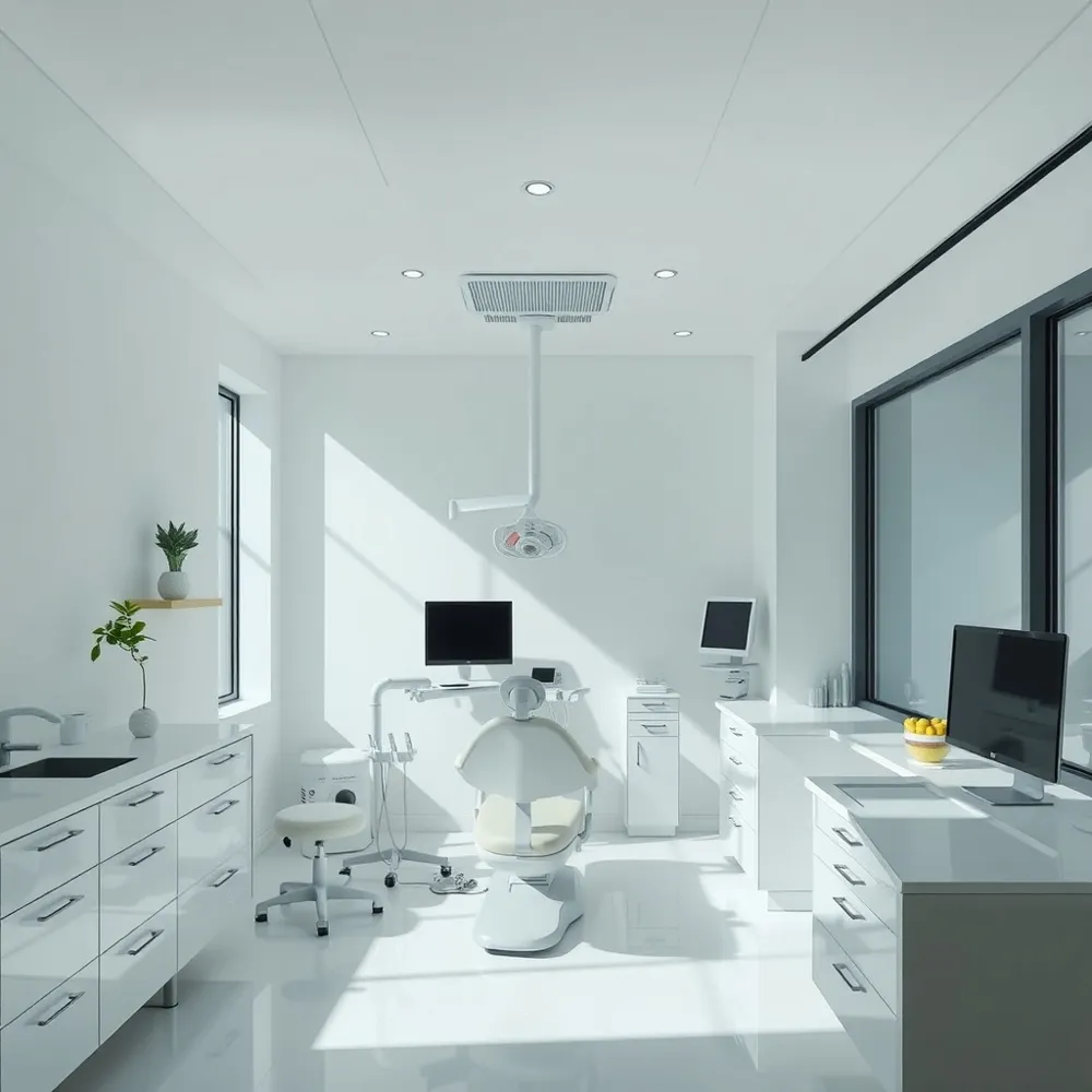
Understanding Digital X-Ray Diagnostics in Early Oral Health Detection
![]()
What is an overview of digital X-ray diagnostics in the early detection of oral conditions?
Digital X-ray diagnostics are a fundamental part of modern dental care, used to identify potential oral health issues at their earliest stages. These advanced imaging techniques create highly detailed, high-resolution images of teeth, bones, and surrounding tissues, which can reveal hidden dental problems such as cavities, bone infections, gum disease, tumors, and other abnormalities. Because digital sensors capture images immediately, dentists can quickly assess and diagnose conditions that might otherwise go unnoticed during regular exams.
Compared to traditional film X-rays, digital radiography reduces patients’ exposure to radiation significantly—by up to 70-90%. This makes the process safer, especially for vulnerable populations like pregnant women. The images are stored electronically, allowing for easy sharing with specialists, monitoring over time, and efficient record-keeping.
Various forms of digital X-rays serve specific diagnostic needs. Bitewing X-rays are ideal for detecting cavities between teeth, while periapical X-rays provide detailed images of individual teeth and their roots. Panoramic X-rays offer a broad view of the entire mouth, including the jaw and sinuses, aiding in comprehensive assessments. Cone beam computed tomography (CBCT) provides three-dimensional imaging for complex cases, such as implant planning or evaluating jawbone structure.
Overall, digital X-ray technology enhances early detection, enabling timely intervention, improving treatment outcomes, and supporting preventive oral healthcare strategies.
Benefits and Advantages of Digital X-Ray Technology for Dental Health Monitoring
![]()
What are the benefits and advantages of digital X-ray technology for dental health monitoring?
Digital X-ray technology has revolutionized dental diagnostics with multiple notable benefits. One of the most significant advantages is safety; digital X-rays reduce radiation exposure by up to 90% compared to traditional film X-rays. This lower radiation level makes the procedure safer for patients, notably children and individuals requiring frequent imaging.
The quality of images captured with digital X-rays is exceptionally high. These high-resolution images can be instantly viewed on a computer screen, allowing dentists to enlarge, adjust contrast, and manipulate the images for better visualization. This clarity supports early detection of issues like cavities, bone infections, gum disease, and other hidden abnormalities.
Efficiency in diagnosis and treatment planning is greatly improved with digital technology. Immediate access to images accelerates decision-making, enables quicker interventions, and enhances patient experience. Digital files are stored electronically, making record-keeping straightforward and enabling seamless sharing with other healthcare professionals, which promotes collaborative care.
Digital X-ray systems are also environmentally friendly, eliminating the need for chemical processing used in traditional film development. This not only reduces chemical waste but also aligns with sustainable practices.
Furthermore, advances in digital imaging include 3D imaging capabilities and AI integration. These innovations improve diagnostic accuracy, especially for complex cases involving detailed assessments of bone density, root structures, and nerve positioning. Overall, digital X-rays contribute significantly to preventative care by supporting early detection and monitoring of oral health conditions, ultimately helping to maintain better overall oral health and quality of life.
Enhancing Early Diagnosis and Intervention of Oral Diseases Through Digital Imaging
![]()
How do digital X-rays aid in the early diagnosis and intervention of oral diseases?
Digital X-rays play a crucial role in early detection of oral health issues. They produce high-resolution images instantly, allowing dentists to identify subtle signs of problems such as cavities, gum disease, and tumors that might not be visible in a routine clinical exam.
Using advanced digital sensors and imaging software, these radiographs enable enhancements like contrast adjustments, zooming, and image manipulation. These features improve the clarity of the images, making it easier to detect early-stage lesions or bone deterioration.
One of the significant advantages of digital X-rays is their safety profile. They reduce radiation exposure by up to 70-90% compared to traditional film X-rays, enabling safer routine screenings.
Further technological developments include digital subtraction radiography, which can highlight changes over time, and 3D imaging with cone-beam computed tomography (CBCT). These tools offer detailed views of dental structures, supporting precise diagnosis and monitoring.
Incorporating digital X-rays into routine care accelerates diagnosis, aids in early intervention, and supports better patient outcomes. This technology not only enhances diagnostic confidence but also facilitates timely treatment planning, preventing more severe oral health issues in the future.
Scientific Evidence Supporting the Effectiveness of Digital X-Ray Technology
![]()
What scientific and research-based evidence supports the effectiveness of digital X-ray technology in dentistry?
Research studies consistently demonstrate that digital X-ray systems deliver diagnostic accuracy on par with traditional film radiography. Multiple studies, including evaluations of CCD Dixi3 and PSP Digora PCT sensors, show similar sensitivity and specificity in detecting early dental issues like non-cavitated approximal caries. Remarkably, digital X-rays are capable of identifying hidden problems such as decay, bone infections, and tumors effectively.
A significant advantage of digital X-rays is their capacity to reduce radiation exposure by up to 80-90%, making them considerably safer for patients. This reduction is achieved through advancements in sensors, software optimization, and radiation reduction features incorporated into modern systems.
Recent technological developments, including cone-beam computed tomography (CBCT) and AI-based analysis, further enhance diagnostic precision. Deep learning models like CNNs (Convolutional Neural Networks) and AI software such as OverJet can automatically detect cavities, periodontal disease, and other anomalies with high accuracy. These AI tools not only facilitate early detection but also support treatment planning and patient education.
Guidelines from professional organizations advocate the adoption of digital modalities for their safety and reliability. They emphasize justified use, appropriate protective measures, and technology standards that promote optimal imaging while safeguarding patient health.
In summary, a robust body of scientific evidence confirms that digital X-ray technology is a reliable, safer, and highly effective tool for early diagnosis and management of various oral health conditions.
Digital vs. Traditional Radiography: A Comparative Analysis
![]()
How does digital radiography compare to traditional radiography methods in the early detection of oral conditions?
Digital radiography provides notable improvements over traditional film-based approaches in detecting oral health issues early on. One of its major advantages is the ability to produce high-resolution images instantly, which allows for quicker diagnosis and prompt treatment planning.
The clarity and detail of digital images enhance the visualization of small or early-stage lesions, such as minor decay or subtle bone changes that might be missed with conventional X-rays. Advanced image processing software further improves diagnostic accuracy by allowing adjustments like zooming and contrast enhancement, helping dentists identify issues that are difficult to detect otherwise.
Safety is another critical benefit. Digital X-rays reduce radiation exposure by up to 80–70% compared to traditional film X-rays. This makes repeated imaging safer for patients while maintaining high image quality.
Moreover, digital images are stored electronically, facilitating easy sharing among dental professionals and integration with artificial intelligence (AI) tools that aid in automated diagnosis. This integration streamlines workflow, supports early detection, and encourages preventive care.
In summary, digital X-ray technology enhances early detection capabilities through superior image quality, improved safety, and seamless digital integration, proving to be more effective than traditional radiography methods in identifying oral conditions at an initial stage.
| Aspect | Traditional X-ray | Digital X-ray | Additional Benefits |
|---|---|---|---|
| Image quality | Good, but limited | High-resolution, adjustable | Enhanced visualization of early lesions |
| Radiation exposure | Higher | Up to 80% lower | Safer for repeated use |
| Speed | Several minutes | Immediate | Faster diagnosis and treatment |
| Storage & sharing | Physical films | Electronic files | Easier sharing, collaboration |
| Integration with AI | Not available | Fully compatible | Supports automated diagnostics |
| Use in early detection | Moderate | Superior | Better prognosis and prevention |
Digital radiography is redefining how early oral conditions are detected, offering a safer, more precise, and efficient diagnostic approach.
Cutting-Edge Advances: Artificial Intelligence and 3D Imaging in Digital X-Ray Diagnostics
What recent advances in digital X-ray technology, including artificial intelligence integration, have improved diagnostic capabilities?
Recent breakthroughs in digital X-ray systems have taken dental diagnostics to new heights. Enhanced sensors now provide high-resolution images that can be seen instantly on monitors, allowing for faster decision-making. These improvements are complemented by the addition of 3D imaging, especially cone beam computed tomography (CBCT), which offers detailed views of teeth, bones, and surrounding tissues.
One of the most transformative developments is the integration of artificial intelligence (AI) algorithms. These AI tools, often based on deep learning models like convolutional neural networks, excel at pattern recognition within complex imaging data. They enable highly accurate detection of abnormalities such as early-stage caries, bone loss, and other lesions.
Several FDA-cleared AI applications, including Overjet’s platform, are now used for automatic caries detection and bone level analysis. These tools interpret radiographs objectively and consistently, reducing diagnostic variability and helping clinicians identify issues before they become clinically obvious.
Beyond just detecting anomalies, AI-driven systems also support workflow enhancement by automating image analysis and report generation. This process improves efficiency, saving time for dental professionals while maintaining a high standard of accuracy.
In summary, the latest advances—ranging from sharper sensors and 3D imaging to intelligent software—are bringing a new level of precision and speed to digital dental diagnostics. They bolster early detection, optimize treatment planning, and streamline practice operations, ultimately contributing to better patient outcomes.
Current Trends and Recommendations in Digital Dental Imaging Practice
What are the current practices, trends, and recommendations in the use of digital dental imaging?
Modern digital dental imaging has become an integral part of routine dental care. It primarily involves digital radiography, which uses digital sensors in place of traditional film to produce high-resolution images instantly displayed on monitors. This technology enhances diagnostic accuracy, allowing practitioners to detect dental decay, bone infections, gum disease, tumors, and other abnormalities early.
Recent trends include the use of cone-beam computed tomography (CBCT), magnetic resonance imaging (MRI), and ultrasound to obtain detailed 3D images of oral structures. These advanced imaging techniques provide comprehensive insights into complex cases such as implant planning and jaw disorders while keeping radiation doses significantly lower—by up to 80% compared to traditional X-rays.
Intraoral scanners and 3D printing are revolutionizing restorative procedures, enabling precise digital impressions and quick fabrication of crowns or bridges. AI integration is another growing trend, with software that analyzes radiographs to identify early signs of disease, automate workflow, and support clinical decision-making. AI systems like Overjet's platform are FDA-cleared for detecting caries and quantifying bone loss.
Further, digital workflows foster collaboration and improve patient engagement, with digital images enabling clearer communication about diagnoses and treatment options. This shift towards digital tools also emphasizes safety; by reducing radiation exposure and improving image clarity, practitioners can provide safer, more effective care.
Recommendations focus on continuous professional training on emerging technologies, investing in AI solutions, and adopting a patient-centered approach. Regular imaging promotes early detection and preventive care, ultimately enhancing oral health outcomes and quality of life.
An understanding of ongoing technological advancements and their integration into daily practice is essential for dental professionals aiming to stay at the forefront of diagnostic precision and patient safety.
| Technology/Trend | Impact | Additional Details |
|---|---|---|
| Digital radiography (X-rays) | Immediate high-resolution images, reduced radiation exposure | Up to 80% less radiation than traditional films |
| CBCT, MRI, ultrasound | Advanced 3D imaging, better diagnostics, lower doses | Useful for complex cases and detailed bone assessment |
| Intraoral scanners & 3D printing | Precise restorations, digital impressions | Speeds up restorative workflows |
| Artificial Intelligence (AI) | Automated detection, workflow support, patient education | AI software highlights anomalies, supports early diagnosis |
| Digital workflows | Better collaboration, record-keeping, patient communication | Enhanced visual clarity and educational tools |
| Digital safety features | Safer imaging, minimized radiation risks | Emphasis on radiation dose reduction |
Adopting these innovative practices ensures a more accurate, safe, and patient-friendly approach to dental diagnostics, aligning with the future of digital dentistry.
The Future of Early Oral Health Detection with Digital X-Ray Technology
Digital X-ray diagnostics have undeniably reshaped the landscape of oral health care by enabling earlier, safer, and more accurate detection of a wide spectrum of oral conditions. Supported by robust scientific evidence and propelled by innovative advancements like artificial intelligence and 3D imaging, digital radiography enhances diagnostic confidence and patient outcomes. Embracing these technologies aligns with current best practices and promotes preventive care strategies that can profoundly improve oral health-related quality of life. As digital imaging continues to evolve, ongoing integration of AI and improvements in sensor technology promise even greater precision and efficiency in dental diagnostics, ultimately supporting better health for patients worldwide.
References
- Dental diagnostics: How do digital X-rays work, and why ...
- Diagnostic Accuracy of Digital and Conventional ...
- Advancements in Digital X-Ray Technology
- Artificial intelligence-produced radiographic enhancements ...
- Digital X-rays: a safer option for monitoring your family's ...
- A visualization system for intelligent diagnosis and ...
- AI Dental Imaging vs Digital X-Rays vs Traditional Imaging
- review digital dentistry and its impact on oral health-related ...
- Artificial intelligence in dental radiology: a narrative review
- ADA Releases Updated Recommendations to Enhance ...