A Closer Look at Dental Imaging
Dental X-rays provide useful views of teeth and surrounding structures, but some questions call for a different perspective. Cone beam computed tomography (CBCT) is a specialized imaging method that produces three-dimensional views of teeth, jawbone, and nearby anatomy. It may help when conventional images do not show enough detail to answer a specific clinical question, such as how a tooth relates to a nerve or the available bone for treatment planning. RadiologyInfo.org’s overview of dental CBCT explains its role and limits.
At Veenstra Family Dental in Midland Park, modern imaging is considered as part of personalized care, not as an automatic step. The practice’s digital imaging approach includes CBCT scans that provide comprehensive images of the jaws and skull to support diagnosis when appropriate.
A scan is not needed for every patient or dental visit. The decision depends on the examination, the information already available, and whether three-dimensional detail is likely to affect diagnosis or treatment planning. The FDA guidance on dental CBCT advises using it when clinically necessary and minimizing exposure. This article explains what the scan can show, how it is performed, and how clinicians weigh its benefits and limitations for each patient.
What a CBCT Scan Shows
 At Veenstra Family Dental, imaging is considered in the context of each patient’s needs. A dentist may recommend a cone beam computed tomography (CBCT) scan when regular dental X-rays do not provide enough information to answer a specific question or plan care.
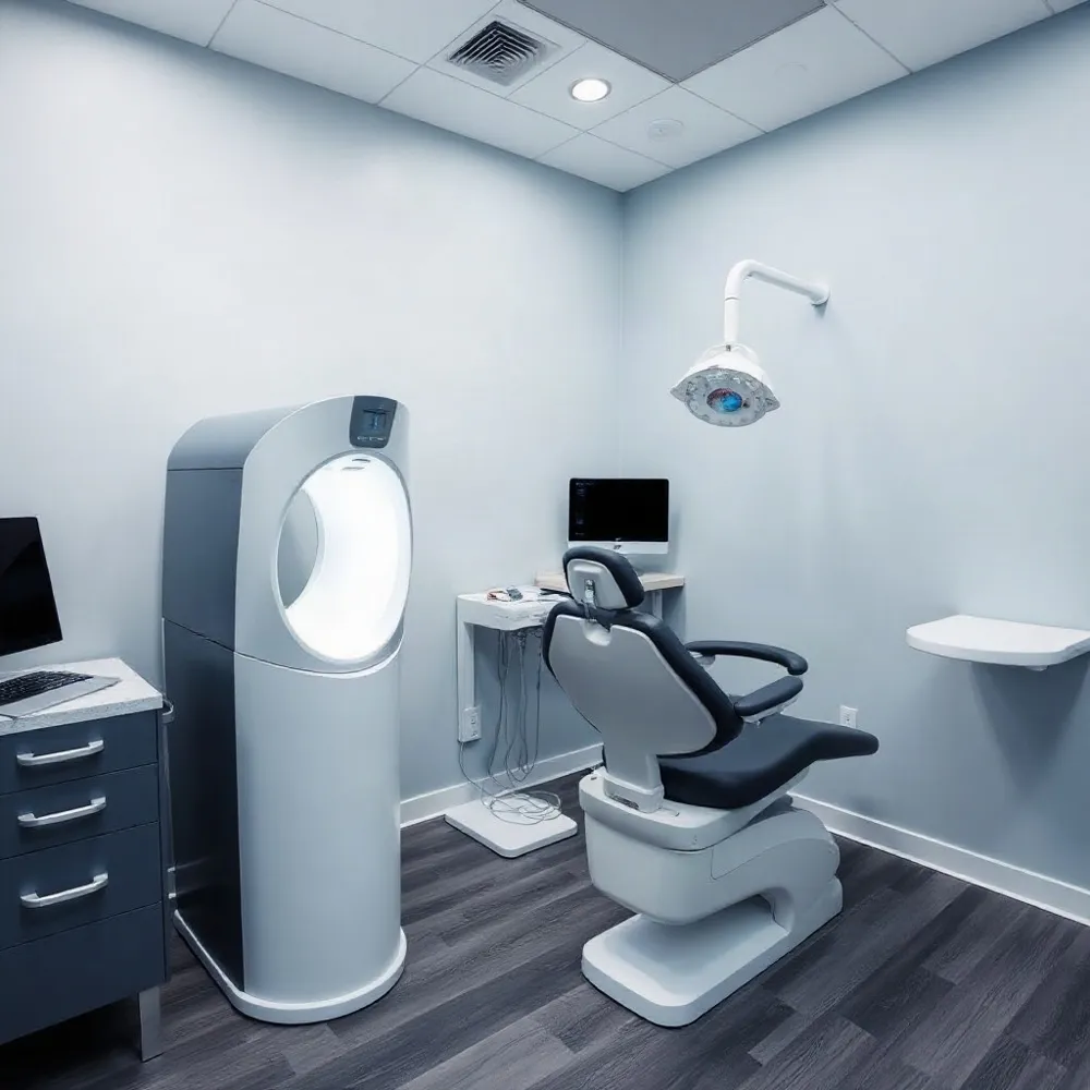
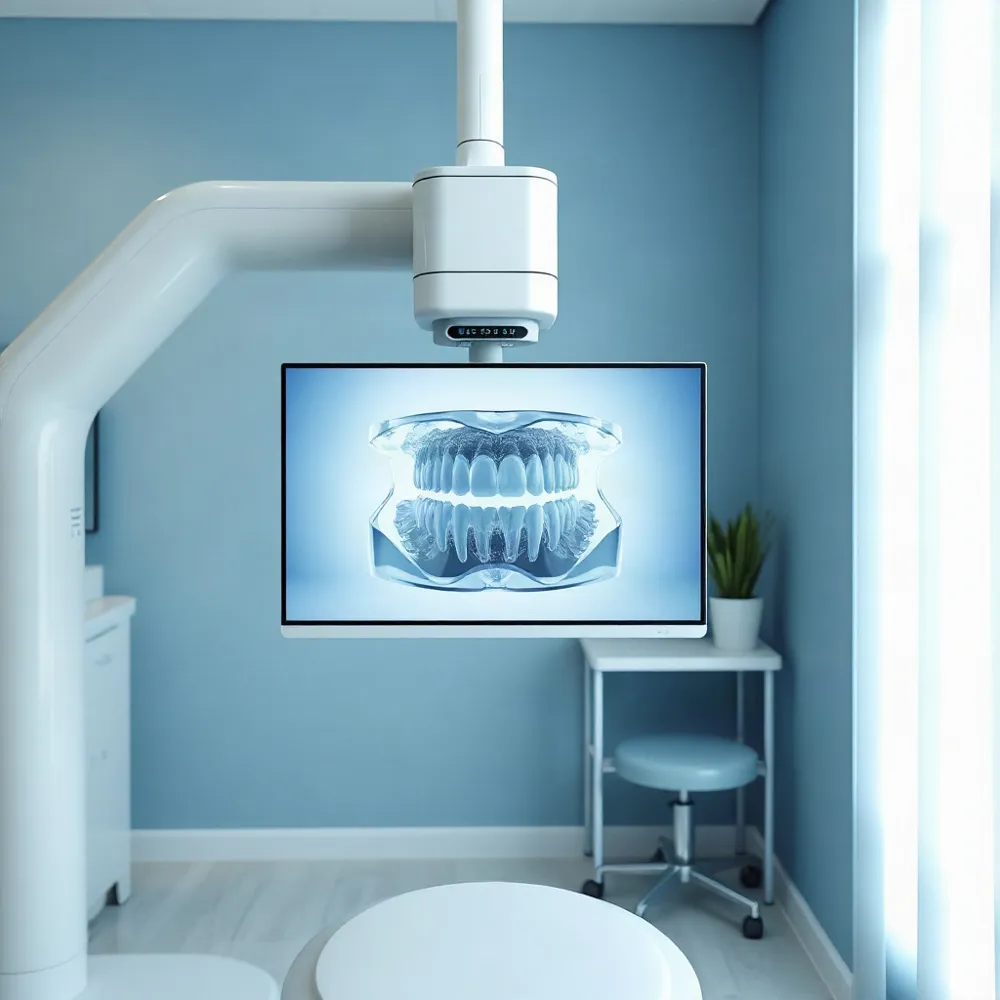
A CBCT scanner rotates a cone-shaped X-ray beam and detector around the head, recording data that software reconstructs into three-dimensional views of the teeth, jaws, and nearby structures, including nerve pathways. These views let a clinician examine anatomy from different directions, rather than relying on a single flat image. RadiologyInfo.org’s overview of dental cone beam CT explains how the scanner’s images are combined into a 3D view.
Conventional dental radiographs remain useful, but their two-dimensional images can make structures overlap or appear distorted. CBCT may clarify relationships that are difficult to judge on those images, such as the position of an impacted tooth near a nerve or the bone available at a potential implant site. The review of CBCT imaging in dentistry describes how three-dimensional views can reduce the effects of superimposition and distortion.
The scan is especially useful for detailed views of teeth and bone, including the jaw and facial bones. It may help assess impacted teeth, jaw concerns, dental injuries, or complex root canal anatomy, and support planning for selected treatments. It is not a routine scan for every visit: the dentist should have a specific clinical question that the added detail can help answer.
CBCT can show some soft tissues, but it does not provide the same soft-tissue detail as conventional medical CT. If muscles, glands, or other soft tissues are the main concern, another imaging method may be more suitable. At Veenstra Family Dental, the purpose of any recommended imaging can be discussed with the dentist, including questions about radiation exposure or possible pregnancy.
How the Scan Is Performed
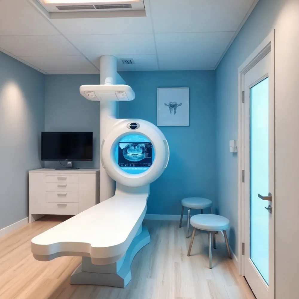 A dental CBCT scan is a brief, noninvasive examination. Depending on the scanner, you may sit or lie down while the dental team positions the area to be imaged. The machine rotates around your head, capturing images from different angles that software combines into a three-dimensional view. You will be asked to stay still during the rotation so movement does not blur the images. You can breathe normally.
How long the scan takes depends on the equipment and the size of the area being imaged. A review reports scanning times of about 5 to 40 seconds, while full-volume scans commonly take 20 to 40 seconds and regional scans may take less than 10 seconds. These are scan times, not the length of the full appointment. RadiologyInfo.org’s CBCT guide describes the procedure and preparation.
Usually, no special preparation is needed. You may be asked to remove jewelry, glasses, hearing aids, or removable dental appliances, so bring appliances with you if requested. Tell the dental team if you may be pregnant or if remaining still could be difficult. At Veenstra Family Dental, imaging is considered in relation to each patient’s needs; the team can explain why a scan is being considered and what to expect. For more on the practice’s approach to digital imaging, see its overview.
Where Three-Dimensional Detail Helps
At Veenstra Family Dental, three-dimensional imaging is considered when a specific clinical question calls for more detail than standard images provide. A CBCT scan is not needed for every patient or procedure; its value depends on whether the added view can help guide diagnosis or planning.
For implant planning, CBCT can show the height and width of the proposed site and the position of nearby anatomy, including the inferior alveolar canal. These views may help a clinician assess where an implant could fit while accounting for surrounding structures. See how three-dimensional detail supports implant planning.
The scan may also clarify the position of an impacted tooth and its relationship to neighboring teeth, nerves, or other important structures when two-dimensional images leave uncertainty. For selected root canal concerns, CBCT can help assess complex root or canal anatomy, a suspected fracture, resorption, or a lesion near the root. It is generally reserved for cases where conventional images do not answer the question, rather than routine endodontic care, as described in this review of CBCT applications in dentistry.
Other possible uses include planning for some orthodontic cases, assessing periodontal bone defects that may be hidden by overlapping structures, and examining bony changes in the jaw or temporomandibular joint. CBCT is strongest for bone detail and does not provide the same soft-tissue information as an MRI. The FDA guidance on dental CBCT recommends using it when the needed clinical information cannot be obtained through other imaging.
For selected procedures, CBCT data can support virtual planning and the preparation of a surgical guide. These tools add information for the dental team, but they do not replace an examination, the patient's history, or professional judgment. At Veenstra Family Dental, imaging is part of a personalized assessment, with the decision based on the patient's needs and the question being addressed.
Balancing Information and Exposure
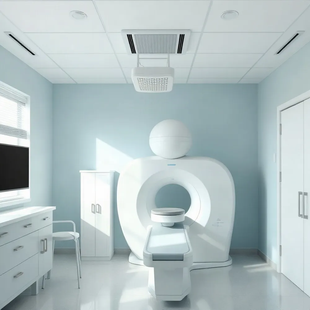 A dental CBCT scan uses ionizing radiation, so it is generally reserved for situations where three-dimensional information may help answer a clinical question. It typically involves more exposure than routine dental X-rays, but less than conventional medical CT. The amount varies with the scanner, exposure settings, and the area imaged, so there is no single dose that applies to every patient. The FDA guidance on dental cone-beam CT recommends using the scan only when needed and minimizing exposure.
A smaller scan area, known as the field of view, can limit exposure to the region relevant to the question. Settings should also be chosen to keep exposure as low as reasonably achievable while still producing images clear enough for diagnosis. A scan set too low may not show the detail the dentist needs. The principles of CBCT field-of-view selection and dose optimization support matching the scan to the clinical need rather than imaging a larger area by default.
Children are more sensitive to radiation than adults, so their age and the reason for imaging should be considered carefully. When a scan is appropriate, child-size protocols and a limited scan area can help reduce exposure. Reviewing earlier images can also help avoid unnecessary repeats. CBCT may reveal unexpected findings that need follow-up, and it is not always the right tool if an examination or another type of image can answer the question.
At Veenstra Family Dental, care is provided for patients across ages, with imaging considered as part of an individual dental assessment. Patients can ask why CBCT is being recommended, whether existing images or an alternative could provide the needed information, what area will be scanned, and how settings will be tailored. This kind of discussion helps clarify the expected benefit, limitations, and exposure before a decision is made. For more on how dental teams weigh imaging information against exposure, see radiation protocols in modern dental imaging.
How Images Are Reviewed
A CBCT scan produces a set of reconstructed views, but the images do not explain themselves. At Veenstra Family Dental, imaging is considered alongside a patient’s symptoms, examination findings, and the clinical question that prompted the scan. This context helps the dental professional determine which details matter for diagnosis or treatment planning.
The reviewer can examine the volume in axial, coronal, and sagittal planes, then use cross-sectional or curved views to assess how teeth, bone, and nearby structures relate to one another. These views can reduce the overlap that may make anatomy harder to judge on a two-dimensional radiograph, as described in this review of CBCT imaging.
Review should cover the full scanned volume, not only the spot of immediate concern. A view of the surrounding anatomy may reveal a finding that deserves follow-up, even if it was not the original reason for imaging. CBCT is especially suited to bony and dental detail, while its soft-tissue limits mean that other assessment may be needed for some questions.
If an image shows something that needs expertise beyond the treating dentist’s scope, the dental team may seek an oral and maxillofacial radiologist’s interpretation or refer the patient to another appropriate specialist. A qualified dentist, oral surgeon, or radiologist can interpret CBCT images, and findings should be explained in relation to the patient’s care, as RadiologyInfo explains.
At Veenstra Family Dental, the aim is to connect imaging findings with the individual treatment plan, rather than treat a scan as a diagnosis on its own. Patients can ask what was seen, whether any follow-up is recommended, and how the result may affect next steps.
Costs and Insurance Questions
How much does a dental CBCT scan cost, and does dental insurance cover it? In the United States, a scan may cost about $150–$600, but this is a general estimate, not a guaranteed price. Costs vary by practice and location, and a limited-area scan may cost less than imaging that covers a larger part of the jaw.
Insurance may cover some or all of the cost when the scan is considered clinically necessary. Coverage depends on your plan and may require clinical documentation or prior approval. Ask Veenstra Family Dental for an estimate and whether the office can check your benefits. Then confirm coverage and any out-of-pocket costs with your insurer before scheduling.
The dentist can explain why the scan is recommended and how its findings may inform your care. For more on planning for dental expenses, see these insurance and cost questions for a first visit.
A Scan Chosen for Your Needs
CBCT can clarify anatomy when ordinary dental images leave a specific question unanswered, but it is not needed for every patient or every visit. The FDA recommends using dental X-rays, including CBCT, only when needed for diagnosis or treatment, with exposure kept as low as reasonably achievable (FDA guidance on dental CBCT).
At Veenstra Family Dental, imaging is considered in the context of each patient’s needs, alongside modern diagnostic tools such as digital imaging. Before a scan, ask what clinical question it is meant to answer, whether another imaging option could help, how the scan area and settings affect exposure, and who will review the images.
You can also ask how the findings may affect treatment choices and what costs or insurance coverage to expect. Sharing records of earlier dental or medical imaging may help the team avoid unnecessary repeat scans. A clear conversation can help you understand the reason for the recommendation and make an informed decision with your dental professional.