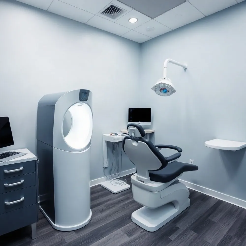
Seeing Beyond the Visual Exam
A visual dental exam can reveal a lot, but it cannot show every surface between teeth or the structures beneath the gums. Dental X-rays create images of teeth and jaws, helping a dentist assess areas such as roots and supporting bone that are not visible during a regular examination. They can also help identify concerns such as decay between teeth or changes in bone that may need further attention.
Digital imaging is a diagnostic aid, not an automatic step at every appointment. The dentist considers your oral health, history, examination findings, and any earlier images before deciding whether new images could help guide care. At Veenstra Family Dental, digital X-rays support a personalized approach to examining and monitoring oral health.
If an image is recommended, the dental team can explain why it may be useful and what it could show. You can ask questions or share concerns before imaging; the American Dental Association’s guidance encourages patients and dentists to discuss recommendations and make informed decisions together.
How Digital Images Are Made
 Dental X-rays, also called radiographs, show teeth and jaw structures that a visual exam cannot fully assess, such as tooth roots and supporting bone. They can help identify decay between teeth or beneath fillings, bone loss, impacted teeth, and signs of infection. Dentists may also use them to plan care or monitor healing, based on a patient’s health history, symptoms, and clinical findings.
With digital imaging, an electronic sensor captures information from the X-ray beam and sends it to a computer, where software displays the image. Film X-rays record the image on photographic film, which must be developed with chemicals. Digital images are usually available soon after capture, so the dental team can review them with you without waiting for film processing. Cleveland Clinic explains how dental X-rays are used and what they can show.
A digital image can be enlarged or adjusted to help a clinician inspect a particular area. These tools can make details easier to examine, but they cannot correct every limitation caused by image quality, positioning, or technique. Images can also be stored electronically, compared with earlier records, and shared with another provider when appropriate. At Veenstra Family Dental, digital X-rays support modern dental care, with image recommendations based on each patient’s needs.
Digital systems may use less radiation than film systems, but a reduction percentage is not a guarantee for every image or patient. Exposure varies with the equipment, settings, and type of image. The American Dental Association’s radiography guidance recommends choosing images according to clinical need and considering prior images when they can answer the question. Ask your dental team what an image is intended to show and how it will inform your care.
Choosing the Right View
 Different images answer different clinical questions, so the right choice depends on what your dentist needs to examine. At Veenstra Family Dental, digital X-rays support care tailored to each patient, from children to adults.
A periapical image is a focused view taken with a sensor inside the mouth. It shows one or a few teeth from crown to root, along with nearby bone, and can help assess a concern such as infection around a root. A panoramic X-ray captures a wider view of the teeth, jaws, and surrounding structures.
Panoramic images can help assess impacted teeth or inform planning for treatment such as braces, extractions, or implants. Because they show a broad area, they provide less detail about an individual tooth than a focused intraoral image. Bitewing images, by contrast, can help reveal decay between teeth and changes near the supporting bone.
Cone beam CT creates a three-dimensional view and may be considered when standard images cannot provide the information needed. It is not a routine substitute for 2D X-rays; the American Dental Association’s imaging guidance recommends choosing imaging according to clinical need. Your dentist can explain which view fits your exam and why.
Personalizing Imaging and Safety
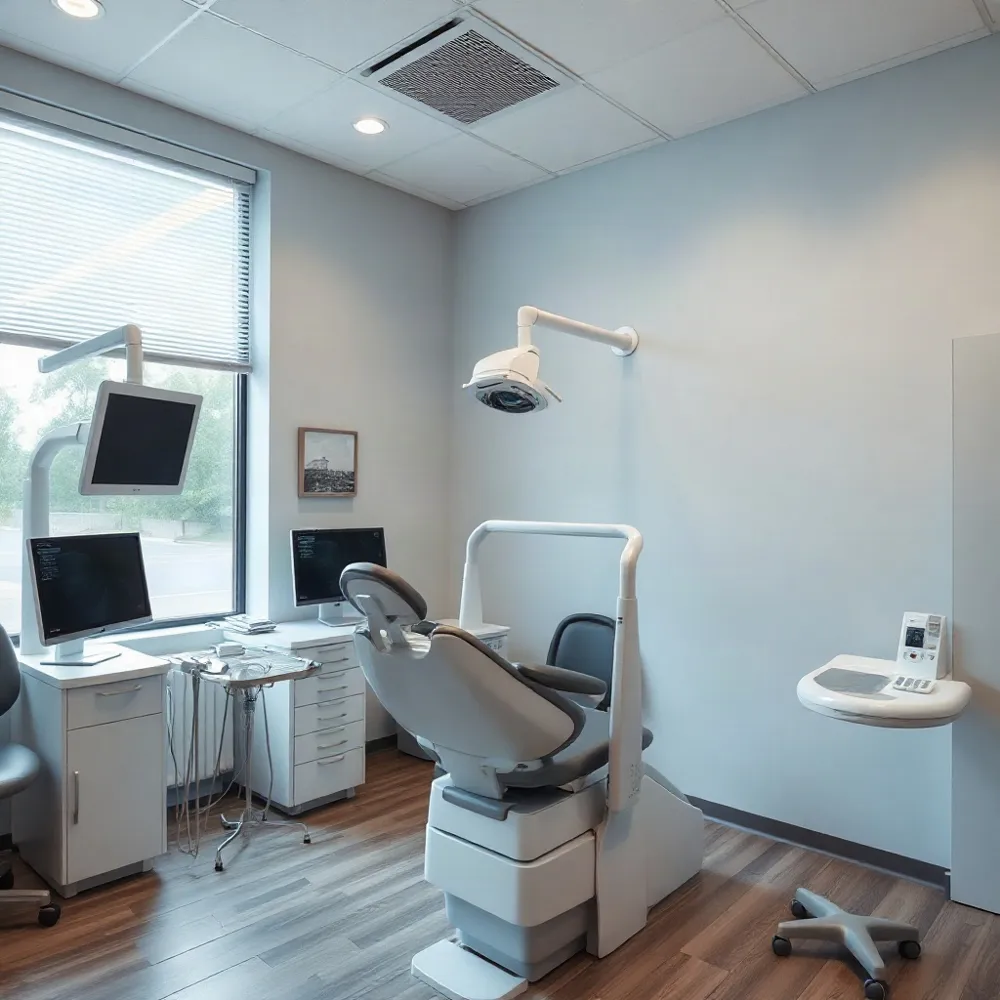 Dental X-rays use a low level of radiation, but exposure is not zero. A dentist recommends an image when its findings may help diagnose a concern or guide care, weighing that benefit against the small potential risk. The American Dental Association’s guidance on dental X-rays emphasizes that images should support, not replace, a clinical examination.
The ADA’s ALARA principle means keeping exposure “as low as reasonably achievable.” In practice, that means choosing images for a clear clinical reason, checking whether suitable images from earlier visits are available, and positioning the patient and limiting the beam carefully. At Veenstra Family Dental, digital X-rays provide images that can be reviewed as part of an individualized examination, rather than treating imaging as a one-size-fits-all step.
How often should I have dental X-rays?
There is no universal schedule. The dentist considers your oral health, age, symptoms, history of cavities or gum problems, risk factors, examination findings, and when you last had images. Someone with stable oral health may need imaging less often than a patient with new symptoms or a history of dental disease. The ADA also advises that X-rays be ordered only when they are expected to provide information that can benefit care.
Are dental X-rays safe, and can I decline them?
Tell your dentist about concerns, including pregnancy or other health circumstances, and ask how a recommended image could affect your care. Digital imaging can offer practical benefits, but it does not remove radiation exposure or make every image necessary. Veenstra Family Dental uses digital X-rays within a personalized dental evaluation; the appropriate view and timing depend on your clinical needs.
You may decline an X-ray. Before deciding, ask what the image is intended to show and what might be missed without it. Decay between teeth, infection, or changes in supporting bone may not be apparent during an examination, and a dentist may need to discuss whether planned treatment can safely proceed without the image. Cleveland Clinic describes dental X-rays as a tool for assessing concerns that may not be visible on visual examination, and supports discussing risks and benefits with your dentist when you have questions about imaging.
Comfort, Records, and Practical Questions
Digital imaging can make records easier to view and share, but the experience still depends on the sensor, positioning, and the reason for the image. At Veenstra Family Dental, digital X-rays are used to help assess concerns such as cavities between teeth, gum disease, and tooth roots, with imaging chosen to support an individual exam.
What are the possible drawbacks of digital dental X-rays?
Some intraoral sensors can feel bulky or uncomfortable, especially for people with a sensitive gag reflex. Positioning and technique also affect image quality. Software may adjust an image for clearer viewing, but it cannot correct every limitation, so a repeat image may sometimes be needed. Digital X-rays still use radiation and should be taken only when clinically appropriate, as the American Dental Association’s radiography guidance explains.
Can I request a copy of my dental X-rays?
Yes. Contact your dental office to ask for a copy and how it can be delivered to you or another provider. Offices may have different request procedures and may charge a reasonable fee for copying or delivery. Veenstra Family Dental’s digital X-ray service supports electronic imaging, but ask the office about its process and any requirements.
How much does a 3D dental X-ray cost?
A cone beam CT scan creates three-dimensional views and uses specialized equipment. It may cost more than a standard image, and fees and insurance coverage vary. Ask the dental office for the scan fee and an estimate of your out-of-pocket cost before proceeding. The decision to use 3D imaging should reflect whether it provides information needed for your care, not simply whether the technology is available; Cleveland Clinic’s overview of dental X-rays describes CBCT as an option often used for implant planning.
A Conversation About Your Images
Digital X-rays can help inform diagnosis and care, but they are one part of the picture. Your dentist’s examination and knowledge of your health history help determine whether imaging is appropriate for you.
If an image is recommended, ask what it is meant to show, whether earlier images can answer the same question, and how the recommendation fits your individual needs. The American Dental Association’s guidance on dental X-rays supports choosing images through discussion and clinical judgment. A clear conversation can help you feel informed about the next step.