A Clearer Look Inside Your Smile
During my years as a dentist, I've taken tens of thousands of X-rays. The moment that changed my perspective came with a young patient who was terrified of the large, intimidating machines we used to rely on. Watching her fear fade when I switched to a small, quiet digital sensor made me realize how much the patient experience matters. That single encounter reshaped how I approach every diagnostic image we capture at our practice, and it's why I'm so committed to the technology we use today.
Traditional film X-rays required longer exposure times and produced images you had to develop with chemicals. Digital systems replace that film with an electronic sensor that captures the image instantly. According to the Cleveland Clinic, digital X-rays use up to 90% less radiation than traditional film, and they produce a higher-resolution image that can be enhanced, zoomed, and shared with you on a chairside monitor. That's a meaningful safety improvement for every patient, especially children and those who need frequent imaging.
But the real shift for me wasn't just about the technology; it was about what it allowed me to do clinically. With instant feedback on the screen, I can spot a cavity between teeth, check the health of the bone around a tooth, or monitor the development of a child's permanent teeth without making anyone wait. A 2018 study in the National Institutes of Health database reviewed the health effects of dental diagnostic X-rays and found that while the doses are low, the cumulative effect of repeated imaging is something dentists should manage carefully. That research deepened my conviction: use the lowest possible dose, but don't sacrifice the diagnostic information that helps me catch problems early.
When I think about how this changed me, it's not about the technology for its own sake. It's about the trust a patient places in me when they open their mouth and let me look. Every time I choose a digital sensor over film, I'm making a choice to reduce their exposure, to show them exactly what I see, and to involve them in their own care. That's the philosophy I bring to every appointment at Veenstra Family Dental, and it's why I'm confident that modern imaging isn't just a convenience, it's a cornerstone of the kind of gentle, transparent dentistry I want to provide.
The Discovery That Changed Medicine
Dental X-rays work by passing a small, controlled beam of radiation through your teeth and gums onto a sensor or film. Dense structures like tooth enamel and bone absorb more of the X-ray energy, so they appear lighter in the resulting image, while softer tissues like gums and nerves let more radiation through and appear darker. This contrast lets your dentist see hidden cavities, bone loss, and other issues that are invisible to the naked eye. If you want the underlying physics explained simply, HowStuffWorks describes the process in an accessible way.
The technology behind these images has advanced considerably. Traditional film X-rays have largely given way to digital radiography, which uses electronic sensors instead of film to capture images in seconds. Digital systems produce higher-resolution images with significantly less radiation exposure, and they allow instant viewing on a screen. According to the Cleveland Clinic, digital X-rays are a common and safe diagnostic tool in modern dentistry. You can also read more about how digital radiography works from a technical standpoint.
The Radiation Question
A common concern is radiation exposure, but the levels involved in dental X-rays are remarkably low. A single set of digital bitewing X-rays exposes you to roughly the same radiation as a short airplane flight or a day spent outdoors in the sun. The American Academy of Oral and Maxillofacial Radiology confirms that dental X-rays are safe when used appropriately, and the benefits of early detection far outweigh the minimal risks. For a deeper look at the health effects, the National Institutes of Health has published research on the topic.
Digital vs. Traditional X-Rays
Digital X-Rays. Use electronic sensors, produce instant high-resolution images, and expose patients to up to 80-90% less radiation than traditional film. They are faster, more comfortable, and easier to store and share electronically.
Traditional Film X-Rays. Require chemical processing, take longer to develop, and use higher radiation levels. They still work, but most modern practices, including ours, have transitioned to digital for safety and efficiency.
At Veenstra Family Dental, we use digital X-rays exclusively. Our advanced imaging tools allow us to detect cavities, bone loss, and other conditions at their earliest, most treatable stages, as explained in our article on digital X-ray diagnostics. This means you spend less time in the chair and get more accurate diagnoses, with a fraction of the radiation you'd receive from older film-based systems.
What to Expect During Your Visit
Getting dental X-rays is quick and painless. Your dental professional will place a small sensor in your mouth and ask you to bite down gently while the image is captured. The entire process usually takes just a few minutes. The images appear instantly on a screen, so we can walk you through what we see and answer any questions right away. If you're curious about the technology behind the scenes, our team's dental radiographer plays a key role in ensuring every image is precise and comfortable.
| Aspect | Traditional Film X-Ray | Digital X-Ray (Veenstra) |
|---|---|---|
| Radiation | Higher | Up to 90% less |
| Time to Image | Minutes | Seconds |
| Image Quality | Good | Excellent, enhanced |
| Comfort | Bulky film | Thin sensor, easy |
| Storage | Physical files | Electronic records |
What Exactly Are X-Rays?
In 1895, Wilhelm Röntgen accidentally discovered X-rays while experimenting with cathode rays in his laboratory. He noticed that a fluorescent screen glowed even when the tube was covered, leading him to realize that a new type of radiation was penetrating the material. Röntgen called these rays "X-rays" to signify their unknown nature. This discovery earned him the first Nobel Prize in Physics in 1901 and laid the foundation for a diagnostic tool that would transform medicine and dentistry.
Early dental X-rays were crude and required long exposure times, sometimes several minutes. Patients experienced significant radiation exposure, and images were captured on glass plates or film. Over the decades, technological advancements have dramatically reduced exposure times while improving image quality. Modern digital radiography, for example, uses electronic sensors instead of film, offering clearer images with significantly less radiation. According to the Cleveland Clinic, digital X-rays are a valuable diagnostic tool that helps dentists detect hidden dental issues that are not visible during a routine exam.
From Film to Digital: The Evolution
The shift from traditional film-based X-rays to digital radiography has been one of the most significant changes in dental diagnostics. Digital systems capture images instantly, allowing for immediate review and faster treatment decisions. Unlike traditional film, digital images can be enhanced, enlarged, and stored electronically. This not only improves diagnostic accuracy but also makes it easier to share images with patients and other specialists. As a result, digital dental X-rays have become the standard of care in modern practices, including ours at Veenstra Family Dental.
Digital X-rays use electronic sensors that are connected to a computer. The sensor is placed in the mouth, and a small amount of radiation is used to capture the image. The data is then transmitted to a computer, where it is displayed on a screen within seconds. This process eliminates the need for chemical processing, which was time-consuming and produced hazardous waste. Furthermore, digital images can be viewed immediately, allowing for real-time patient education and collaborative treatment planning. For a deeper understanding of how this technology works, the experts at TWI explain the principles of digital radiography in detail.
One of the most significant advantages of digital X-rays is their ability to aid in the early detection of oral conditions. Conditions such as cavities, bone loss, and impacted teeth can be identified at their earliest, most treatable stages. This is particularly important because many dental problems are invisible to the naked eye and only become noticeable once they have progressed. Early detection not only improves treatment outcomes but also reduces the need for more invasive and costly procedures. The American Dental Association and the Cleveland Clinic both emphasize the role of X-rays in diagnosing hidden issues that a visual exam might miss.
Radiation Safety: A Priority in Modern Dentistry
Patient safety is a top concern when it comes to dental X-rays. While all radiation exposure carries some risk, the amount used in dental imaging is extremely low, especially with modern digital technology. Digital X-rays use up to 90% less radiation than traditional film X-rays. Lead aprons and thyroid collars are still used to minimize exposure to the rest of the body, although many modern digital systems require even less protection. The American Academy of Ophthalmology and other health organizations provide guidance on the safety of dental X-rays, and leading dental practices like ours follow rigorous protocols to ensure patient safety.
The amount of radiation from a single dental X-ray is comparable to the background radiation we are exposed to daily from natural sources. To put this in perspective, a set of four bitewing X-rays exposes a patient to about 5 microsieverts of radiation, which is less than the 10 microsieverts one receives from a cross-country flight. This is why dental organizations, including the American Dental Association, consider dental X-rays safe for routine use, especially when proper precautions are taken. At Veenstra Family Dental, we use digital X-rays to minimize radiation exposure while maximizing diagnostic accuracy, ensuring our patients receive the highest standard of care.
The Future of Dental Imaging
As technology continues to evolve, so does the field of dental imaging. Cone beam computed tomography (CBCT) is a three-dimensional imaging technique that provides detailed views of the teeth, jaws, and facial structures. This technology is particularly useful for planning dental implants, evaluating impacted teeth, and diagnosing TMJ disorders. Another emerging trend is the use of artificial intelligence (AI) to analyze dental images. AI algorithms can detect patterns and anomalies that might be missed by the human eye, improving diagnostic accuracy and efficiency. These advancements are making dental care more precise, comfortable, and personalized for patients.
At Veenstra Family Dental, we are committed to staying at the forefront of dental technology. Our practice uses advanced digital imaging tools to provide our patients with accurate diagnoses and comfortable experiences. We believe that a clear understanding of the technology behind your dental care empowers you to make informed decisions about your health. Whether you are coming in for a routine checkup or a more complex procedure, you can trust that our team uses the latest and safest imaging techniques to ensure the best possible outcomes.
How an X-Ray Machine Creates an Image
When you sit for a dental X-ray, the action begins in a small glass tube that generates the radiation used to capture your teeth. Understanding how an X-ray tube works, from the physics of the cathode and anode to the role of the tungsten target, helps explain why modern digital systems deliver clearer images with lower exposure. For a deeper look at how this translates into your care, see how digital X-ray diagnostics support early detection of oral conditions.
The Cathode and Anode: The Heart of the Tube
Inside every dental X-ray tube, a heated filament acts as the cathode, releasing a cloud of electrons when electricity passes through it. These electrons accelerate toward a positively charged metal disk called the anode, which contains a small tungsten target. When the high-speed electrons collide with the tungsten, most of their energy converts to heat, but a small fraction produces X-ray photons. This process, known as bremsstrahlung, is the same fundamental physics described in resources on how X-ray radiography works from physics to safety.
From Photons to Your Dental Image
Once generated, the X-ray photons pass through your teeth and surrounding tissues onto a sensor or film. Dense structures like enamel and bone absorb more photons, appearing lighter, while cavities and infections allow more photons through, showing up as dark areas. This contrast is what lets your dentist spot problems early. Digital sensors capture this information instantly, offering advantages over traditional film, as outlined in a comparison of digital dental X-rays versus traditional methods.
Safety and Precision in Modern Dental Radiography
Modern dental X-ray systems are engineered to minimize radiation while maximizing diagnostic quality. Digital radiography uses faster sensors and lower exposure settings, reducing radiation by up to 80% compared to film, according to the Cleveland Clinic’s guide to dental X-rays. This efficiency means less time in the chair and greater peace of mind for patients of all ages. At Veenstra Family Dental, we pair these advanced imaging tools with a gentle approach, ensuring every image is captured comfortably and accurately.
| Component | Function | Diagnostic Benefit |
|---|---|---|
| Cathode | Emits electrons via heated filament | Controls exposure time |
| Anode | Accelerates electrons, contains tungsten target | Produces X-ray photons efficiently |
| Digital sensor | Captures photons as electronic data | Immediate images, lower radiation |
The physics behind X-ray tubes may seem complex, but the result is straightforward: clearer, faster, and safer dental imaging. If you’re curious about how this technology benefits your next visit, our team can explain the process in plain terms, showing you exactly what we see and why it matters for your oral health.
Digital Radiography in Dentistry
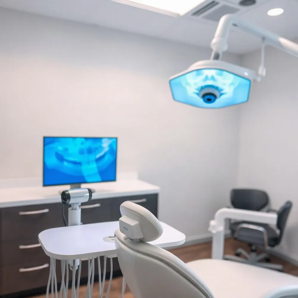 Digital dental X-rays replace traditional photographic film with an electronic sensor placed inside your mouth. When the dentist or dental radiographer activates the X-ray unit, photons pass through your teeth and surrounding tissues before striking the sensor. The sensor converts those photons into an electrical signal, which a computer then translates into a detailed on-screen image. The entire process takes just seconds, and the image appears instantly on the monitor.
The Technology Behind the Image
At the heart of this system is a solid-state sensor, typically a charge-coupled device (CCD) or a complementary metal-oxide-semiconductor (CMOS) chip. When X-ray photons hit the sensor, they free electrons in the silicon, creating a charge pattern that mirrors the density of the structures the beam passed through. A computer reads this pattern and reconstructs it as a grayscale digital image. This process is fundamentally different from film-based radiography, where the image is stored on a light-sensitive sheet that must be developed chemically. You can read more about the physics behind the technology in this guide to how digital radiography works.
The result is a highly detailed, instantly viewable image that reveals cavities, bone loss, infections, and other oral conditions at their earliest stages. This early-detection capability is one of the main reasons digital X-ray diagnostics have become a standard of care in modern dentistry.
Digital vs. Traditional Radiography
The shift from film to digital sensors brings several measurable advantages for patients. Traditional film requires chemical processing, which takes time and introduces potential for errors. Digital imaging eliminates those steps entirely, producing a usable image in real time. The Cleveland Clinic explains the different types of dental X-rays and notes that digital systems use up to 90% less radiation than traditional film-based methods.
Another difference lies in the image itself. Digital sensors have a wider dynamic range, meaning they can capture both very dark and very light areas in a single exposure. This reduces the need for retakes and allows the dentist to enhance contrast, zoom in on specific areas, and measure structures with greater precision. These capabilities translate directly into more accurate diagnoses and, ultimately, better treatment outcomes for you.
Radiation Safety and Exposure
One of the most common questions patients ask is whether digital X-rays are safe. The answer is yes. According to the American Academy of Implant Dentistry, the radiation exposure from a single dental X-ray is extremely low, equivalent to a few days of natural background radiation. Digital systems reduce this exposure further because they require less radiation to produce a diagnostic image compared to film.
A 2018 study published in the Journal of the American Dental Association reviewed the health effects of dental diagnostic X-rays and concluded that the benefits of early detection far outweigh the minimal risks for most patients. The study emphasized that modern digital techniques, combined with proper shielding and guidelines, keep exposure well within safe limits. For more details on how radiation exposure compares to other everyday sources, see this comparison of digital dental X-rays to other radiation sources.
What to Expect During Your Visit
When you sit in the chair at Veenstra Family Dental, a dental radiographer positions a small sensor holder in your mouth to capture the necessary views. The sensor is connected to a computer by a thin wire, and each image takes only a second or two to acquire. You won't experience any discomfort, and you won't have to wait for film to develop. Within moments, your dentist reviews the images on a monitor and discusses the findings with you, making the entire appointment more efficient and transparent.
The process is designed with your comfort in mind. Because the sensor is smaller and more flexible than traditional film packets, many patients find it more comfortable. And because the images appear instantly, there's no need for retakes due to underexposure or processing errors, which shortens your time in the chair. For a closer look at the technology behind this experience, explore how modern digital imaging improves treatment plans.
The Benefits at a Glance
| Aspect | Digital X-Ray | Traditional Film X-Ray |
|---|---|---|
| Image time | Seconds | Several minutes |
| Radiation dose | Up to 90% lower | Standard |
| Chemical processing | None | Required |
| Image enhancement | Yes, easy | Limited |
| Retakes due to errors | Rare | Common |
| Storage & sharing | Digital, instant | Physical files |
Reading the Shades: Why Bone Looks White
 When you hear the word "X-ray," concern about radiation exposure is natural. But dental X-rays are among the most thoroughly studied and regulated procedures in medicine. The amount of radiation in a single digital dental X-ray is remarkably low, and the protective measures we take at Veenstra Family Dental ensure that your exposure stays as minimal as possible.
Digital X-rays use a fraction of the radiation of traditional film-based imaging, and they provide sharper, more accurate images. As the Cleveland Clinic explains, these images help dentists detect cavities, bone loss, infections, and other conditions that are invisible to the naked eye. The scientific evidence on dental X-ray safety indicates that the benefits of early detection far outweigh the minimal risks, especially when modern digital technology is used.
At Veenstra Family Dental, we take your safety personally. Our team uses lead aprons and thyroid collars for every X-ray, and we follow the American Dental Association's guidelines on imaging frequency. We never take X-rays "just because" — every image is clinically justified and tailored to your individual needs. This is what sets us apart from practices that may rely on outdated film X-rays: our commitment to advanced technology means less radiation and more precise diagnostics for you and your family.
Radiation Exposure: The Facts
When you sit back in the dental chair and the X-ray sensor is positioned, the resulting image is far more than a shadow on a screen. Dental X-rays use a small, focused beam of radiation to create a detailed picture of your teeth, roots, and jawbone. This image lets your dentist spot issues that a visual exam simply cannot reach, such as early-stage decay between teeth, bone loss, infections, or impacted teeth. According to the Cleveland Clinic, dental X-rays are a standard diagnostic tool that can reveal hidden dental problems, including abscesses and cysts, before they become painful or costly to treat.
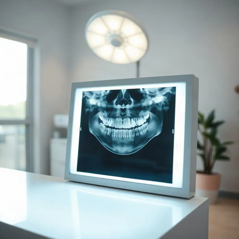
The process is quick and precise. A digital sensor or film is placed inside your mouth, and a carefully aimed beam passes through your teeth and gums. Dense structures like enamel and bone absorb more of the beam and appear lighter, while softer tissues let more radiation through and show up darker. This contrast is what creates the familiar black-and-white image your dentist reads. The underlying physics has been understood for over a century, as explained by HowStuffWorks, but modern digital systems have refined the process dramatically.
How Much Radiation Are We Really Talking About?
A common worry is radiation exposure, but the numbers are reassuring. A full set of digital dental X-rays delivers roughly the same amount of radiation you would receive from a short airplane flight or a day spent outdoors in the sun. In fact, dental digital radiography exposes you to far less radiation than traditional film-based X-rays, often by as much as 80% to 90% less, according to the American Academy of Implant Dentistry. To put it in perspective, the average person receives about 3 millisieverts (mSv) of background radiation each year from natural sources like the sun and soil; a single dental X-ray is typically around 0.005 mSv, which is less than 1% of your annual background dose.
That low dose is no accident. Modern digital systems use sensors that are highly sensitive to X-rays, meaning less radiation is needed to produce a clear image. As noted by the Cleveland Clinic, the amount of radiation from a dental X-ray is extremely low, and the benefits of early detection far outweigh the minimal risk. For patients who are still concerned, your dentist can use lead aprons and thyroid collars for extra protection, and the practice follows strict guidelines set by organizations like the American Dental Association.
From Pixels to Diagnosis: What the Image Reveals
Once the digital image is captured, it appears on a computer screen within seconds. This immediacy is a huge advantage over traditional film, which requires chemical processing and a waiting period. Digital X-rays allow your dentist to zoom in, adjust contrast, and measure the exact size of a cavity or the depth of a bone pocket. This level of detail supports earlier intervention, which often means smaller fillings, less invasive procedures, and a better long-term outcome for your smile.
The technology behind the scene is straightforward. A digital sensor converts X-ray photons into an electrical signal, which is then translated into a pixel-based image on a computer. As the folks at TWI explain, digital radiography captures the image in a fraction of a second, reducing the chance of blur from patient movement. This is especially helpful for children or patients with a strong gag reflex, as the shorter exposure time means less discomfort.
Why It Matters for Your Next Visit
Understanding what your dentist is looking at during your appointment can turn anxiety into confidence. When you know that the small sensor is helping to catch a developing cavity before it reaches the nerve, or to monitor the health of a previous root canal, it is easier to appreciate the value of the image. For a family practice like Veenstra Family Dental, this technology means more personalized, proactive care for every member of your family, from the youngest to the most senior.
If you are curious about how these images are used to plan treatments, or if you want to see what your own X-rays reveal, the next time you are in the chair, ask. The image is yours, and your dentist can walk you through it in plain language. That transparency is part of what makes modern dental care feel less like a mystery and more like a partnership. And with the low radiation exposure, the peace of mind is well earned.
Types of Dental X-Rays and When They're Used
Dental X-rays fall into two broad categories: intraoral, where the sensor or film is placed inside your mouth, and extraoral, where the sensor stays outside. Each category is designed to capture specific views of your teeth, jaws, and surrounding structures.
Intraoral X-Rays: Detailed Views of Individual Teeth
Bitewing. Shows the crowns of the upper and lower teeth in a single area of the mouth. Bitewing X-rays are the standard tool for detecting cavities between teeth and assessing the bone level around teeth, which can indicate gum disease.
Periapical. Captures the entire tooth, from the crown down to the root tip and the surrounding bone. This type is used to detect root structure abnormalities, abscesses (infections), and to monitor healing after procedures like root canals.
Occlusal. Reveals the full arch of teeth in the upper or lower jaw in one image, helping to locate impacted teeth, jaw fractures, or other issues on the floor of the mouth or the roof of the mouth.
Extraoral X-Rays: Big-Picture Views of the Jaw and Skull
Panoramic. Provides a single, wide image of the entire mouth, including all teeth, both jaws, the temporomandibular joints (TMJ), and the sinuses. It's used for planning implant placement, detecting impacted wisdom teeth, and evaluating jaw problems or tumors.
Cephalometric. Captures a side view of the head, showing the teeth in relation to the jaw and facial profile. Orthodontists rely on this view for treatment planning to correct bites and align teeth.
Cone Beam CT (CBCT). Generates a 3D image of the teeth, jaws, nerves, and sinuses. This advanced scan is invaluable for complex procedures like dental implant placement, diagnosing difficult root canal anatomy, and oral surgery planning.
In orthodontics, X-rays are essential for seeing beneath the gum's surface. They allow the orthodontist to assess tooth position and development, evaluate jaw growth, check for missing or extra teeth, and monitor root health, all of which are critical for planning safe, effective treatment.
There is no one-size-fits-all X-ray schedule. At veenstradental.com, the need for X-rays is determined by a clinical exam rather than a calendar date. For patients with healthy teeth and gums, a set of bitewing X-rays may be sufficient every 12 to 18 months. Those with a history of cavities, gum disease, or ongoing restorative work may need them more frequently to monitor changes.
How Often Should You Have X-Rays?
 For healthy adults with no active dental concerns, most general guidelines recommend new X-rays every 12 to 24 months. This aligns with the standard checkup interval for many patients and is sufficient to monitor for early signs of decay or gum disease between cleanings.
Children, teens, and patients with a history of frequent cavities or active gum disease often need a shorter interval — typically every 6 to 12 months. Growing mouths change faster, and children may need more frequent imaging during periods of rapid tooth loss or crowding. At Veenstra Family Dental, we tailor the schedule to each patient's clinical needs rather than applying a one-size-fits-all rule.
Your dentist will also factor in personal dental history, the presence of restorations, past treatment outcomes, and any symptoms you may be experiencing. A patient with healthy tissue and no recent cavities may be comfortable at the 18-month mark, while someone with recurring issues or a complex treatment plan might benefit from more frequent monitoring.
Your specific schedule is best determined during a conversation at your next visit. Many modern practices, including ours, rely on modern digital X-ray diagnostics to keep image data current while minimizing radiation exposure, making it safe to capture updates only when clinically warranted.
A Safe, Personalized Approach to Imaging
Stepping into the imaging room for the first time can feel unfamiliar, but a modern dental X-ray is a quick, comfortable process. Most visits follow the same simple flow: you sit in the chair, a protective lead apron is placed over your chest and lap, and a small sensor or film holder is positioned inside your mouth. The dentist or dental radiographer steps behind a shield to capture the images, and each exposure takes just a fraction of a second. The entire series is usually complete within a few minutes, and you can get back to your day immediately afterward.
At Veenstra Family Dental, the process is designed with patient comfort in mind. The team uses digital X-ray systems that eliminate the wait for film development, so your images appear on a screen almost instantly. This means less time with your mouth open and fewer chances for the uncomfortable gag reflex that some patients associate with older film-based methods.
A common worry is radiation exposure, and it is fair to ask about it. The amount used in a digital dental X-ray is remarkably low. According to the American Academy of Implant Dentistry, a full set of digital dental X-rays delivers a similar dose to what you would receive from a short airplane flight or a day in the sun. When compared to traditional film, digital sensors reduce radiation exposure by up to 80%, as noted by Cleveland Clinic.
Modern technology also means the images are not only safer but more useful. Digital radiography produces high-resolution pictures that can be enhanced and zoomed, allowing your dentist to spot small cavities, bone loss, or early infections that might be missed on film. This aligns with the broader movement toward gentle, precise dentistry that minimizes discomfort while maximizing diagnostic accuracy.
Addressing Common Questions and Concerns
Patients often wonder how often they really need X-rays. The answer depends on your individual risk, but for most people, a full set of bitewing images is recommended every 12 to 18 months as part of a routine checkup. This frequency allows your dentist to monitor changes and catch issues early, when they are easiest and least expensive to treat.
Another frequent question is whether X-rays are safe during pregnancy. The short answer is that dentists take extra precautions, using lead aprons and thyroid collars to minimize any exposure. If you are pregnant or suspect you might be, let your dentist know at the start of your appointment so they can adjust the imaging plan accordingly.
If you still have concerns about the process, you are not alone. Many people feel anxious about the unknown. The best way to ease that anxiety is to talk openly with your dental team. They can explain each step, show you the equipment, and answer any questions you have before the first image is taken. This transparency is part of the patient-focused care that makes a visit to a modern dental practice feel less like a procedure and more like a conversation.
Rest assured that the technology used today is a far cry from the bulky, high-radiation machines of the past. With digital sensors, advanced software, and a team that prioritizes your comfort, a dental X-ray is a safe, fast, and painless step toward a healthier smile.